Health and healing
Medical history, medicines, smoking, previous gum disease and factors such as diabetes can affect healing and risk.

Preparing bone for implant treatment
Additional surgery is sometimes considered when the available jawbone cannot support an implant in the planned restorative position
Bone grafting does not create an automatic route to implant treatment. The need for grafting, the type of procedure and whether an implant can be placed at the same appointment depend on the anatomy, health of the site, intended tooth position and individual healing factors.
The purpose of grafting
After a tooth is removed, the surrounding ridge commonly changes in height and width. Gum disease, infection, trauma, previous surgery and natural anatomy can also leave insufficient bone for the intended implant.
The aim of grafting is to create or preserve a volume of bone that supports an implant in a position compatible with the final crown, bridge or denture. It may also help support the gum contour, but no graft can guarantee a particular aesthetic or functional result.
Diagnosis before surgery
Medical history, medicines, smoking, previous gum disease and factors such as diabetes can affect healing and risk.
Clinical examination assesses ridge shape, gum health, restorative space and whether active infection must be treated first.
Radiographs and, when justified, CBCT imaging relate the planned tooth to the available bone, sinus, nasal cavity and other anatomical structures.
Different defects require different approaches
Graft material may be placed when a tooth is removed or around an implant where a local gap or exposed surface requires support. Implant placement may be simultaneous or delayed according to site conditions and stability.
Particulate graft material is protected with a barrier membrane so that bone-forming tissues can occupy the planned space. The wound must remain stable and well covered during healing.
A larger width or height deficiency may require grafting before implant placement. This increases treatment time and may require another surgical appointment after maturation.
More extensive deficiencies may require a block graft or bone collected from another site. This carries additional donor-site considerations and is not necessary for most routine implant sites.
The upper back jaw
The maxillary sinuses are air-filled spaces above the upper premolar and molar teeth. When there is insufficient bone beneath a sinus for the intended implant, its lining may be carefully elevated to create a protected space for graft material and new bone formation.
A sinus lift may be performed before implant placement or at the same appointment. The decision depends particularly on the residual bone, the required augmentation and whether an implant can obtain adequate initial stability.
Surgical access
The sinus floor is approached through the implant preparation site. It is generally considered where a more limited increase is required and sufficient existing bone allows simultaneous implant placement. The technique is less extensive, but it is not risk-free and is not appropriate for every sinus.
A small access window is prepared from the side of the upper jaw. The sinus lining is elevated and graft material placed beneath it. An implant may be placed simultaneously when conditions allow; otherwise, the graft is allowed to mature before a later implant procedure.
Consented clinical example
These radiographs show the same patient and upper posterior treatment site. The pre-operative image informed assessment of the available bone beneath the maxillary sinus. The definitive implant-supported crown was fitted approximately seven months after that assessment, following the planned surgical and restorative stages.
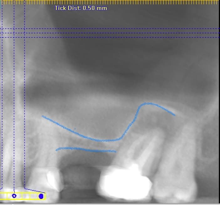
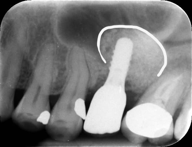
The images were taken with different radiographic projections and magnification. They provide clinical context, but they should not be treated as a directly scaled measurement comparison or as a guarantee of the result another patient might achieve.
The maintenance plan included periodontal maintenance at approximately three-month intervals, a dental examination at approximately six-month intervals, continued monitoring of the tissues, bite and restoration, and a protective bite guard. Recall intervals remain subject to each patient's clinical findings.
Published with the patient's consent. Radiographs form only one part of assessment and follow-up; treatment suitability and outcomes vary between patients.
Informed material choice
Depending on the defect, grafting may use a patient's own bone, processed human donor material, animal-derived material, synthetic substitutes, or a combination. A resorbable or non-resorbable membrane may be used to protect the site.
These materials act primarily as a scaffold while healing occurs; they are not all biologically identical. Their origin, benefits, limitations and any personal, ethical or religious concerns should be discussed before consent. Platelet-rich fibrin may sometimes be used as an adjunct, but it does not replace sound surgical principles or guarantee faster bone formation.
A staged biological process
Swelling, bruising, discomfort and minor bleeding can occur after grafting. A graft commonly requires several months to mature, and the complete interval to a finished restoration may extend beyond six months. Timing varies with the procedure, graft material, implant stability and individual healing. Temporary tooth arrangements should be planned before surgery where appearance or function could otherwise be affected.
Medication and aftercare are prescribed for the individual procedure and medical history. Antibiotics, steroids or decongestants are not presented as a universal routine for every patient.
Possible problems include pain, swelling, bruising, bleeding, infection, wound opening, graft exposure, loss of graft material, inadequate bone formation and failure of an implant to integrate. Further grafting or a change in the restorative plan may be required.
Sinus procedures also carry a risk of tearing the sinus lining. A small perforation may sometimes be repaired during surgery; a larger defect may require the procedure to be stopped and repeated after healing. Sinus infection, nosebleeds, altered sensation and a persistent communication between the mouth and sinus are less common but important complications.
Risk can be higher with smoking, uncontrolled gum disease, inadequate plaque control and certain medical conditions. Individual risks and warning symptoms must be explained as part of consent.
Balanced treatment planning
Alternatives may include a shorter implant where adequate bone remains, placing implants in a different position, a reduced dental arch, an implant-supported removable denture, a tooth-supported bridge, a conventional denture, or accepting the space. Each changes the balance of surgery, cleaning, biting forces, cost and maintenance.
The appropriate comparison is not simply “graft or no graft”. It is which overall treatment plan provides a maintainable restoration with proportionate biological risk.
Documented clinical context
See how local grafting and sinus augmentation formed part of a wider six-implant restorative plan.
View the All-on-6 caseSee a local graft used as one part of managing a traumatised front-tooth site.
View the single-tooth caseSee why removal, grafting and staged reassessment may be required when an existing implant fails.
View the complication caseAuthor and clinical reviewer
DDS · MSc (Conservative Dentistry, Eastman Dental Institute, UCL) · LDS RCS (Eng) · MFDS RCSEd. Dr Daoudi is registered with the General Dental Council as a dentist and Specialist in Prosthodontics, GDC number 78666.
Review Dr Daoudi's qualifications and experience · Search the GDC register
References were checked during the clinical review on 12 September 2026. This page will be reviewed earlier than the scheduled date if material clinical guidance changes.
This page provides general information and does not replace an individual examination, diagnosis or consent discussion.