
01
Assessment of the Failing Anterior Bridge
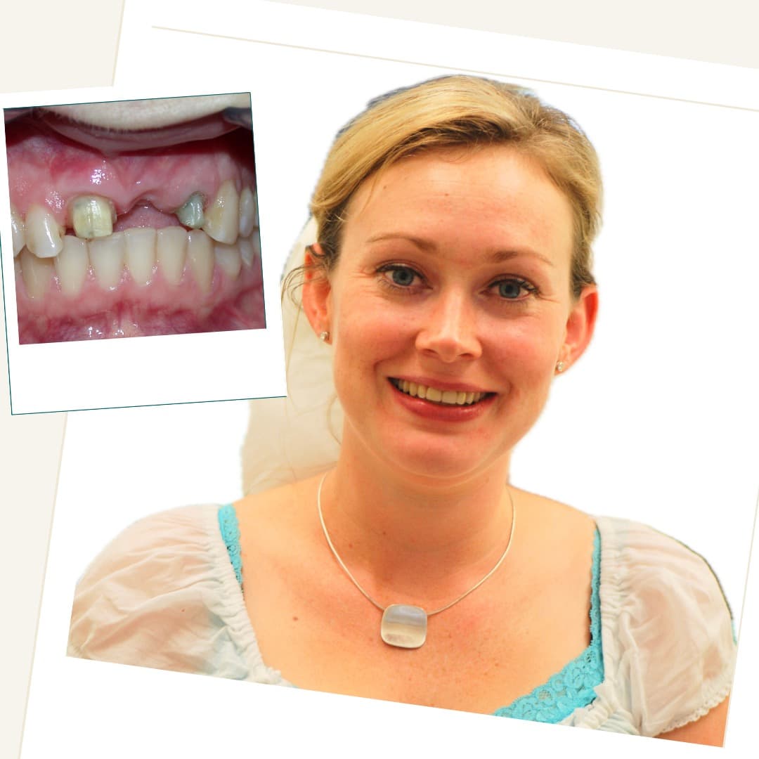
The upper-left lateral incisor had a root fracture beneath the existing restoration, affecting its ability to support the bridge. The upper-right central incisor showed progressive root resorption.
The history, clinical findings and imaging were considered together. As neither abutment tooth offered a predictable restorative foundation, removal and implant-supported replacement were discussed alongside the relevant alternatives, stages and risks.
02
Stage 1: Extractions and Socket Preservation
The compromised upper-right central and upper-left lateral incisors were carefully removed. The extraction sites and remaining facial bone were assessed during surgery.
Socket preservation was used to help limit the expected dimensional changes that follow extraction and to support the later implant plan. It cannot prevent all bone remodelling.
Bone-graft material and platelet-rich fibrin (PRF), prepared from the patient's own blood, were placed where indicated. The sites were then monitored during an approximately four-month healing period.
03
Stage 2: Implant Placement and Guided Bone Regeneration
After the initial healing phase, the anterior ridge was reassessed and two dental implants were placed in positions planned to support the bridge.
Guided bone regeneration was carried out at the same visit where additional facial bone volume was required. The graft combined locally collected autogenous bone with a bovine-derived bone substitute.
A resorbable membrane was placed over the graft to separate it from the overlying soft tissues during early healing. A further healing period of approximately four months was planned before definitive restoration.
04
Stage 3: Implant-Supported Porcelain Bridge
After implant integration and soft-tissue healing had been assessed, the provisional restoration was removed and the implant foundations and restorative space were reviewed.
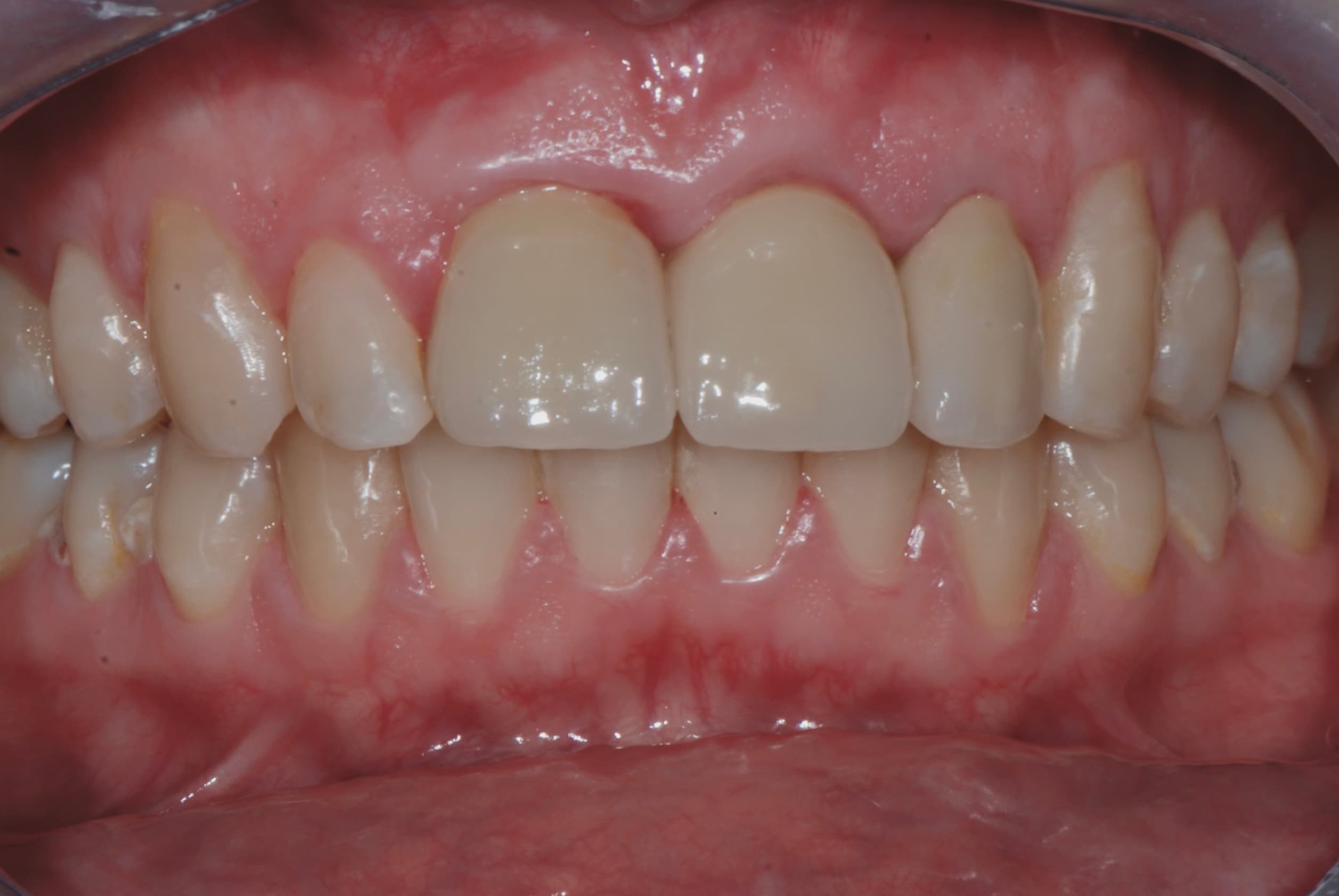
An individually made implant-supported porcelain bridge was fitted. Its fit, cleaning access, shade, contours, speech and bite contacts were evaluated before completion.
05
Stage 4: Maintenance and Continuing Care
The patient received individual guidance on cleaning beneath the bridge and around the implant components, together with a recommended programme of dental and professional hygiene reviews.
Ongoing assessment is important for monitoring the surrounding tissues, ceramic surfaces, bridge fit, screw connections and bite forces. Referred patients can return to their general dentist with a treatment report and maintenance recommendations.