For referring dentists

Complex Endodontic and Tooth-Preservation Referrals

Microscope-assisted assessment and treatment of selected complex cases

Dr Firas Daoudi is registered by the GDC as a Specialist in Prosthodontics and has extensive clinical experience and a particular interest in endodontics. Referrals are welcomed for selected cases involving previous treatment, complex anatomy, restorative complications or uncertainty about whether a natural tooth can be retained.

Collaborative care

Working within the agreed referral remit

The clinical question, proposed scope of care and restorative responsibilities are clarified before treatment. The referring dentist is updated with relevant findings and treatment information, and the patient is returned for continuing care as appropriate.

Radiographs and relevant clinical details can be uploaded through the secure referral form. Please avoid sending confidential patient information by ordinary email.

Consented clinical examples

Selected Complex Endodontic Cases

These anonymised cases illustrate clinical reasoning and technical challenges. They do not promise the same result for another tooth or patient.

Case 1

Root-canal retreatment and perforation repair

An upper lateral incisor supporting a bridge had previously received root-canal treatment and a post. Assessment identified a lateral mid-root perforation and persistent disease associated with the tooth.
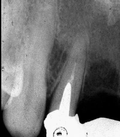
Pre-treatment dental radiograph of an upper lateral incisor bridge abutment with an existing post and root filling
Before treatment: the previously root-treated upper lateral incisor was serving as a bridge abutment and contained an existing post.
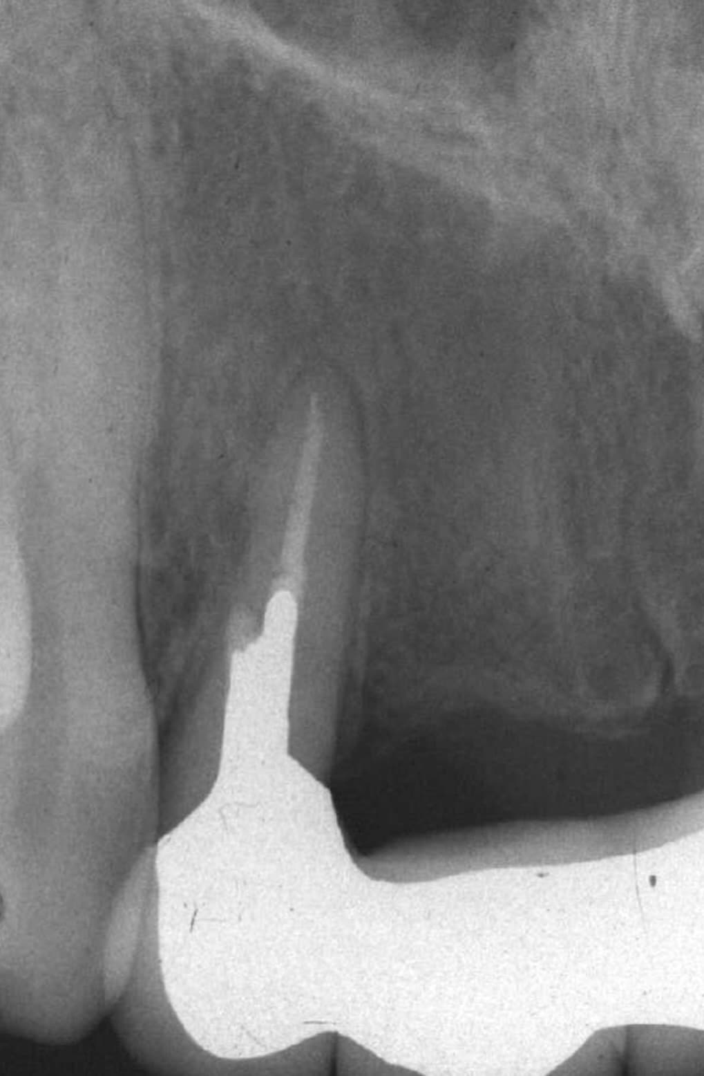
Twenty-year review radiograph after root-canal retreatment and perforation repair of an upper lateral incisor
Twenty-year review: maintained radiographic healing following retreatment, microscope-assisted perforation repair and definitive restoration.

Clinical approach

  • The existing bridge and post were carefully removed under rubber-dam isolation.
  • Previous root-filling material was removed and the canal system was retreated using an operating microscope.
  • The perforation was identified under magnification and repaired using mineral trioxide aggregate (MTA).
  • The tooth was restored with a cast gold post-and-core and a new porcelain-fused-to-metal bridge.
Interpretation and review

The 20-year review radiograph supplied for this case shows maintained periapical bone healing with no evident recurrent radiographic abnormality at the reviewed site. Long-term survival in one case does not predict the outcome of treatment for another tooth.
Case 2

Non-surgical treatment of a large apical radiolucency

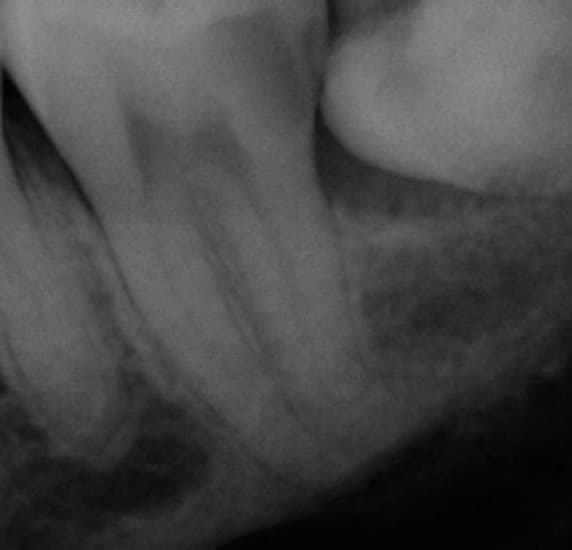
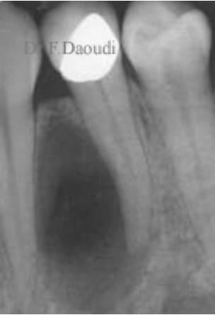
A lower-left first premolar presented with a substantial radiolucent area around the root apex. The aim was to disinfect the root-canal system non-surgically and retain the tooth where clinically appropriate.

Pre-treatment radiograph of a lower-left first premolar with a large apical radiolucency
Before treatment: a substantial radiolucent area is visible around the apex of the lower-left first premolar.
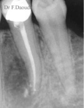
Follow-up radiograph after non-surgical root-canal treatment of a lower-left first premolar
Follow-up: radiographic bone fill is visible after staged non-surgical root-canal treatment.

Clinical approach

  • Treatment was completed in two carefully controlled stages.
  • An inter-appointment intracanal medicament was used between visits.
  • Copious irrigation using a multi-irrigant protocol supported cleaning of the canal system before obturation.
Interpretation and review

The follow-up radiograph demonstrates substantial radiographic bone fill at the previously radiolucent site. Radiographic change must be interpreted alongside symptoms, clinical examination and continued review.

Case 3

Complex root anatomy in a lower second molar

A lower-left second molar presented with markedly curved distal root anatomy. Negotiating, cleaning and filling such anatomy requires detailed assessment, controlled instrumentation and careful working-length management.

Pre-treatment radiograph of a lower-left second molar with pronounced distal root curvature
Before treatment: the lower-left second molar shows pronounced distal root curvature.
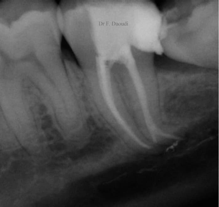
Post-treatment radiograph of a lower-left second molar showing obturated curved root-canal anatomy
After treatment: the obturated canal follows the marked distal curvature, with additional filled lateral anatomy visible radiographically.

Clinical approach

  • The distal root displayed a pronounced apical curvature approaching 90 degrees.
  • The canal system was prepared and obturated while respecting the original root anatomy.
  • The postoperative radiograph also records filled accessory or lateral canal anatomy.
Interpretation and review

This case illustrates anatomical complexity rather than a universal treatment pathway. Curvature, canal diameter, calcification, infection and remaining tooth structure all influence feasibility and risk.

Case selection, risks and alternatives

Root-canal treatment and retreatment cannot be guaranteed. Existing restorations, obstruction, perforation, fracture, resorption, root curvature, infection and the amount of remaining tooth tissue can influence feasibility and prognosis. Following assessment, the options may include monitoring, non-surgical treatment, surgical endodontics, restorative intervention or extraction and replacement.

Clinical review

Reviewed by: Dr Firas Daoudi, GDC 78666, Specialist in Prosthodontics

Last reviewed: 13 September 2026

Clinical images are anonymised and published with patient consent. The page provides general referral information and does not replace case-specific clinical assessment.

Professional referrals

Discuss or Refer a Complex Case

Use the secure referral form to provide the clinical question, relevant history and available radiographs.